
![]()
|
|
|
|
MolDyn |
 RUN program by the command

   ../$MDYN07HOME/mDynQ07 -i inProtcol  -c inPDB [-mdR mdRestXYZVin] [-mv moveRes]
             [-r1 inRestrainA1 ] [-r2 inRestrainA2] [-rB rigBodyFile]
             [-sa saProtocol] [-mn molName]  [-mdX mdFinalPDB] -o runOutFile [-er errorFile]

in parenthesis [ ] are uxilarry files. The auxilary files will be used by program if the main command file
defines respective task.

Command line DESCRIPTION:

-i inProtcol       : file MdynPar.inp  defines protocol for the mDyn particular Run

-c inPDB           : file of the initial molecular structure as molec.pdb file in the PDB format

-mdR mdRestXYZVin  : XYZ+Velocity file to REstart MD from the last snapshot file XYZV , see exaple t5
                     1arb.mdXYZVfin0001.pdb it is USED with $mdRestart keyword in command file
                     inProtcol
                     NOTE!  the initial XYZ will be taken from mdRestXYZVin file !
                           the PDB file inPDB is not USED with the key -mdR

-r1 inRestrainA1   : file defines  of positional restraints for atoms of the  molecule
-r2 inRestrainA2   : file defines atom-atom distance restraints
-rB rigBodyFile    : file defines rigid body segments of the main chain of protein
-mv moveRes        : file defines List of moving Residues
-sa saProtocol     : file defines simulated annealing protocol
-mn molName        : character set defining molecula name.  molName. will be attached to RESULT files
-o runOutFile      : run output file
-mdX mdFinalPDB    : final PDB file of the Energy/MD optimization

Current status of program run is printed on the standart output device (consol) or
can be redirected to user defined file or can be defined in the argument line:
-er errorFile      : error message file : they are dublicated in the runOutFile
#
if file name definition in the argument line is missing for a file
than the default name is used for this file

NOTE! if the command line does not include a key -X , while the command file defines task which need data file coupled with -X keyword,  than program try to find default (standart) name data file in the current directory.
Default names:
#
inProtcol   = ./MdynPar.inp
inPDB       = ./molec.pdb
mdRestXYZVfile = ./mdXYZVin.pdb
moveRes     = ./moveRes.inp
inRestrainA1  = ./restrAt1.inp'
inRestrainA2  = ./restrAt2.inp'
rigBodyFile = ./rigBody.inp
saProtocol  = ./SAprotocol.inp
molName     = space
runOutFile  = ./mDynSB.out
errorFile   = ./mDynSB.err
mdFinalPDB  = ./molMdFin.pdb
#
inProtcol   = ./MdynPar.inp

The nain command file consist of lines with command keyword.
Keyword start with $ sign in the first position of line
One Keyword in line

#example of MdynPar.inp file and keyword description
# MdynPar.inp
$OUTfull                                 ! full extended output of program run

#Initial PDB data quality
$Hread                                   ! read INPUT pdb file with Hydrogens
                                         ! by default OUTshort option is ON
# DEfinition of OPtimized segments of protein:
$fullProtMD                              ! full molecule is flexible
#$MovingRes                              ! defines List of opimized segments

#FORCE FIELD MODIFICATIONS:
#
$shake=2                                 ! all valence bobds are fixed by shake method

$zeroRot                                 ! exclude translation and rotation of the molecule
                                           as rigid body

$hBond128 = 2.0                          ! scaling coeff for H-bonds
                                         ! default=1.0 it is standart force field

$harmAt1PosRst=0.25                      !invoke restraintsA1 type =
                                          positional harmonic restraints for atom position
                                          with harmConst (kcal/A^2).
                                          program need a special file -r1 restrA1File
                                          which defines restrained segmants of protein
                                          see additional description

$distRestrA2                             !invoke restraintsA2 type atom-atom distances
                                          for user defined pairs of atoms in the file
                                          -r2 restrA2File (see additional description)


$rigBody                                 !invoke optimization with frozen internal structure of
                                          protein main chain for user defined segments of sequence
                                          need file -rB rigidBodySegment (see additional description)

$compactForce = 0.5                      ! invoke additional compactization forces
                                         ! to accelerate protein folding
#
$aSoftCore = 0.5                         !invoke SOFTNES for the van der waals atom-atom potential
                                         ! at the small (contact) atom-atom distances
                                         ! Use of the softCore VDW potential helps to optimize
                                         ! BAD molecular structures with many spartial atom-atom clashes
                                         ! values range  0 - 1 from very Soft to standart VDW
#SOLVATION MODEL
$SolvMod = GShell
#
#
# OPIMIZATION PROTOCOL:
$engCalc                               ! do energy calculation
$engOptim                              ! do energy optimization by local Optimizer
$nOptStep=1                            !max N optim steps
#
#PROTOCOL for Molecular Dynamics:
$doMDyn                                ! do MolDynamics
$MDSA                                  !do MolecularDynamis SimAnnealing
                                        needs SAprotocolFile -sa saProtocol File,
                                        see additional description
#
#PROTOCOL of MD equilibration:
#
$initMDTemp=50.00                      !initial Temperature to start MolDyn
$bathMDTemp=50.00                      !thermostat temperature of thermostat i.e. target temperature
$runMDnstep=2000                       !number of time-steps for MD simulation
$mdTimeStep=0.002
#
$NTV=1                                ! MD ensemble definition
#
#
# MD Trajectory writing:
$nwtra=500
$WRpdb                                   ! write snarshort structures in the PDB format
                                         ! default WRpdbq OPTion is ON : extended PDB format
                                         ! PDB + Qatom
#
END
#
NOTE that parameter file formatted, i.e. $ sign should be  the firs character of the line
----------------------------------------------------------------------------
KEYWORD LIST:
        keyw = 'OUTfull'
        keyw = 'WRpdb'
        keyw = 'Hread'
        keyw = 'fullProtMD'
        keyw = 'MovingRes'
        keyw = 'LigRes'
        keyw = 'doLigDock'
        keyw = 'MDSA'
        keyw = 'SolvMod'
        keyw = 'zeroRot'
        keyw = 'hBond128'
        keyw = 'harmAt1PosRst'
        keyw = 'distRestrA2'
        keyw = 'compactForce'
        keyw = 'shake'
        keyw = 'engCalc'
        keyw = 'engOptim'
        keyw = 'nOptStep'
        keyw = 'aSoftCore'
        keyw = 'initMDTemp'
        keyw = 'bathMDTemp'
        keyw = 'mdTimeStep'
        keyw = 'runMDnstep'
        keyw = 'doMDyn'
        keyw = 'mdRestart'
        keyw = 'NTV'
        keyw = 'nwtra'
-----------------------------------------------------------------------------
KEYWORD DESCRIPTION:

#OUTPUT DETAILES:
$OUTfull                                 ! full extended output of program run
                                         ! by default OUTshort option is ON
#
# INPUT PDB FILE DETAILES:
$Hread    ! defines that all Hydrogens will be read from input molecule structure -c inPDB   file
            otherwise the ALL HYDrogens will be restored by the program, i.e.
            all H atoms will be deleted and added according to molecular topology for RESidues.
            Using Library in the ./dat/h_add.dat
NOTE! it is recommended start to works with a new protein without option $Hread even if the PDB
file has all hydrogen atoms, because the hydrogen atom names for protein side chains
have multiple definition in the PDB data base.
It is better if mDyn  program will add all hydrogens to the heavy atoms.

#DEFINITION OF OPTIMIZED RESIDUES:

$fullProtMD                             !defines FULL (i.e. ALL atoms) of the USER molecule
                                         will be free to move in energy relaxation or molDyn

$MovingRes              ! logical keyWord defines that only a defined set of RESidue are free
                 this keyWord is coupled with file -mv moveRes in the argument line to start
                 the program
                 default name for moveRes file is ./moveRes.inp

#EXAMPLE of ./moveRes.inp
#1arb
aaaaaaIIIIiiii
#
MOVRES   1  10       !line defines first and last resudue of moving segments integers devided by space
MOVRES  45  76
MOVRES 115 260
end                  !end or END should be last line if the file
************

#FORCE FIELD DEFINITION:

$hBond128 = 2.0                          ! scaling coeff for H-bonds

$aSoftCore = 0.5                         !invoke van der waals atom-atom potential with modified
                                         ! SoftCore at the small (contact) atom-atom distances
                                         ! SoftCore modification is used for energyOPtimization
                                           and MD equilibration stages.
                                         ! Use of the softCore VDW potential helps to optimize
                                         ! BAD structures with many starical atom-atom clashes
                                         ! values range  0 - 1 from very Soft to standart VDW

$harmAt1PosRst=0.25  ! digital keyWord define RESidue segments with 1 atom position harmonic
                       restrants.
                       0.25 = harmonic restrain Constant K
                       restrEnergy = 0.5*K(r - r0)**2,
                       the reference position r0 = initialXYZinput.pdb - positions from
                       the initial INPut PDB file which defines INItial structure of molecule

    this keyWord is coupled with file -r1 inRestrainA1 of the argument line to start
                 the program mdyn
                 default name for inRestrain file is ./restrAt1.inp

#EXAMPLE  of inRestrainA1 file:
#harmonically restrained RESidue segments
#xxxxxIIIIiiiiaaAAA
#(6x,2i4,a40)
RESTA1   1  63  PBB           ! line starts from keyWord RESTAT numbers=first/last residue of segment
                              ! PBB (only protein backbone atoms are restrained, i.e. side chains are free)
RESTA1  78 120  ALL           ! ALL (all atoms are restrained)
                              ! integers and words are devided by space
end
# ---------------------------------------------------
$distRestrA2                  ! defines optimization/MD with atom-atom dist RestrainA2
                              ! needs file  [-r2 inRestrainA2] in command line
-r2 inRestrainA2 : default name : restrAt2.inp
#
EXAMPLE of inRestrainA2 file:
#harmonically restrained Atom-Atom distances
#xxxxxx
#keyword atom1       atom2       distA HarmConst(kcal/mol*A^2)
RESTA2   ND2  ASN 222 : OG1 THR 219 = 7.0   1.5
RESTA2   O  GLY 170 : OG1 THR 219 = 8.0   2.5
RESTA2   OH TYR 109 : OG1 THR 111 = 7.5   3.0
END
#----------------------------------------------------
$rigBody                    !defines optimizatiom/MD considering some segments of the main chain
                            ! as a rigid body.
                            ! The List of rigid  segments of the main chain is user defined.
                            ! Each segment will keep rigid internal structure of the protein main chain,
                            ! has rotatational and translational degrees of freedom.
                            ! The side chains of the rigid segments are flexible.
#Needs file rigidBody.inp
#EXAMPLE of rigidBody.inp file:
#
RIGB01  11  16       !line defines first and last resudue of moving segments integers devided by space
RIGB02  47  59
RIGB03  77  99
end                  !end or END should be last line if the file
# - - - - - - - - - - - - - - - - - - - - - - - - -
$compactForce = 0.25        ! define additional compactization forces for protein atoms
                            ! Recomended forceParameter = 0.1 - 1.0
# --------------------------------------------------

$shake=2    ! invoke shake subroutine to keep bonds fixed. =1 -bonds with Hydrogen, =2 all bonds

----------------------------------------------------
#Defining of the SOLVation model:
there are 4 variants  of Implicit models
          1 variant of Explicit model
#:
$SolvMod = GShell           ! implicit  Gaussian Shell solvation model
$SolvMod = GShell + WBrg    ! implicit  Gaussian Shell solvation model + WaterBridges between polar atoms
                         ! WaterBridges descride solvent mediated interactions trough stong bound water
                         ! molecules via implicit model of water bridges

$SolvMod = GBorn            ! implicit Generalized Born model + SAS HydroPhobic solvation
$SolvMod = GBorn + WBrg     ! implicit Generalized Born model + SAS HydroPhobic solvation + WaterBridges

$Solv = ExWshell 4.5 [A] ! explicit water shell of 4.5 Angst around protein;
                         ! recomended thikness 3.0 - 6.0 A
---------------------------------------------------
$mdRestart    ! restart molDynamics from a snapshot [molName.]mdXYZVfin000N.pdb
                the file [molName.]mdXYZVfin000N.pdb should be copied to the file mdyn Restart file
                mdXYZVin.pdb

$doMDyn       ! do molecular dynamics
$MDSA         ! do Molecular Dynamical Simulated Annealing
              ! coupled with file -sa SAprotocol which define protocol of the simulated annealing

#EXAMPLE of Aprotocol.inp  file
#SA protocol
#nSAstep 2
#(f10.1,1x,f8.1,1x,3(f6.1,1x)
#      nMDstep    tempTg   SCvdW wfHb128BB wfhB128BS
SAPROT 100000      500.0     0.8    1.0     1.0        !line starts from keyword SAPROT
SAPROT 100000      100.0     1.0    1.0     1.0
END
#
   nMDstep - number of md timeStep
   tempTg  - target temperature in K, this temperature will be reach during ntimeMX steps
   SCvdW   - parameter 0 - 1 to define softness of the van der waals potential. Soft potential
             modifies Potential Energy Surface and decrease  barriers of conformational transitions
   wfHb128BB,
   wfhB128BS - (1 - 0) scaling factors for BackBone-BackBone and
                BackBone-SideChain Hydrogen Bond energy
#--------------------------------------------------
#
# OPIMIZATION PROTOCOL:
$engCalc                               ! do energy calculation
$engOptim                              ! do energy optimization by local Optimizer
$nOptStep=1                            !max N optim steps
#
#PROTOCOL for Molecular Dynamics:
$doMDyn                                ! do MolDynamics
$MDSA                                  !do MolecularDynamis SimAnnealing
                                        needs SAprotocolFile -sa saProtocol File,

#MD EQUILIBRATION:
$initMDTemp=50.00                    !defines initial temperature to start MD
                                     ! recommended low temperature < 50K
                                     ! temperature can be steadelly increased to the 300K and higher
                                     ! USING $MDSA option
$bathMDTemp=50.00                    ! bath temperature in the MD equilibration run
$runMDnstep=2000                     ! number of MD time steps  in the equilibration run
$mdTimeStep=0.002                    ! value of the MD time step in ps,
                                     ! recomended 0.001 - 0.002
$NTV=1                               ! ansemble NTV=0/1
                                     ! =1 md run with constant T
#MD TRAJECTORY WRITING
$nwtra=500                           ! structure XYZ (snapshot) will be written
                                     !as a series of molMdResXXXX.pdb files

$WRpdb                                   ! write snapshort structures in the PDB format
                                         ! default is WRpdbq OPTion is ON : extended PDB format
                                         ! PDB + Qatom column
#* * * * * * * * * * * * * * * * * * * * * * * * * * * * * * * * * * * * * * * * * * * * * * *
#
-c inPDB   file  - standart pdb file

#EXAMPLE of inPDB   file:
****************************************************************************************
NOTE! it is recommended to start to work with a new protein without option $Hread even if the PDB
file has all hydrogen atoms, because the hydrogen atom names for protein side chains
have multiple definition in the PDB data. It is better if mDyn  program will add all hydrogens
to the heavy atoms.
*******************************************************************************************
REMARK: PDB:
ATOM      1  N   GLY A   1      11.726 -10.369  10.598
ATOM      2  H1  GLY A   1      11.921 -11.015   9.807
ATOM      3  H2  GLY A   1      12.518 -10.395  11.271
ATOM      4  H3  GLY A   1      10.852 -10.663  11.079
ATOM      5  CA  GLY A   1      11.567  -9.015  10.090
ATOM      6  HA2 GLY A   1      10.772  -8.977   9.420
ATOM      7  HA3 GLY A   1      12.439  -8.710   9.612
ATOM      8  C   GLY A   1      11.280  -8.099  11.303
ATOM      9  O   GLY A   1      11.256  -8.584  12.493
ATOM     10  N   VAL A   2      11.060  -6.876  11.020
ATOM     11  H   VAL A   2      11.066  -6.574  10.025
etc.
TER                 ! CHAIN TERmination
ATOM   1302  N   GLY A  94      10.957 -15.678  12.832
ATOM   1303  H   GLY A  94      10.735 -14.663  12.877
ATOM   1303  H   GLY A  94      10.735 -14.663  12.877
ATOM   1304  CA  GLY A  94      10.193 -16.559  11.950
ATOM   1305  HA2 GLY A  94       9.428 -16.004  11.516
ATOM   1306  HA3 GLY A  94       9.784 -17.323  12.525
ATOM   1307  C   GLY A  94      11.016 -17.184  10.843
...
etc.
TER                 ! CHAIN TERmination
END                 ! file  END
* * * * * * * * * * * * * * * * * * * * * * * * * * * * * *
#
# PDB mDyn trajectory file description:
#
	Program mDyn generate a series of snapshot files, e.g.,  1arb.molMdRes0nnn.pdb (test/t4)
the molMdResXXXX.pdb file (see example) contains all atomic coordinates and additional information
in the REMARK: lines
####
REMARK: Md result : MdTime(ps):    2.4940
REMARK: $nstep:      1247
REMARK: $nRecPDB:       5
REMARK: RMSD(x0):     0.43   <- RMSD all atom
REMARK: badBond: n,erAv(A)  :     0  0.000    <- number and error Average for bond length in Angstrem
REMARK: badAng : n,erAv(grd):     8   9.42    <- number and error Average for bond angles in grad
# ENERGY TERMS for the given structure
REMARK: $ENERGY:     :Kcal
REMARK: eVbondDef:     100.89315      <-bond deformation energy
REMARK: eVangDef :     441.63705      <-angle deformation energy
REMARK: eImpDef  :      35.68147      <-Improper torsion agle [planarity] energy
REMARK: eTorsDef :     691.25769      <-torsion potentioal energy
REMARK: engVDWR1 :   -1031.16211      <- van der waals energy for cutoff R1=8 A
REMARK: ehBHxY128:    -608.70599      <- H-bondinds energy
REMARK: engCOULR1:    -816.25323      <- COULOMBIC for distances < cutoff R1
REMARK: engCOULR2:      -4.47208      <- COULOMBIC for distances Rij,  R1< rij
To run Ligand docking modules, the main command file MdynPar.inp
have to include the next keywords:

# keywords=value
$LigRes= 282 283          !define start/end ligandResidues

in the inPDB file
                             [(i4,1x,i4) format after= ]
                  !the residues numbers are the same as it is in the initial
                  !inPDB file [united pdb file of protein + ligand]

$doLigDock=1      !run docking for USER defined initial position of Ligand
                  ! as it is in the initial inPDB file [united pdb file of protein + ligand]
                  ! Docking is done via simulated annealing molDynamics
                  ! with coupled temperature and force field variation.
                  ! Ligand CMass can move in vicinity of initial
                  ! position +/- 4.0 A
                  ! Orientational global optimization are done via
                  ! simulated annealing MD with multiple start
                  ! orientations. Initial orientations are uniformly
                 ! cover all orientational phase space with distance = 90 deg

$doLigDock=2        ! run ab initio docking
                    ! This option will seach all binding sites on the
                 ! protein molecular surface including cavities and crevices.
                 ! 1) search of surface cavities, crevicies and groovs
                 ! 2) calculation and scoring of binding site candidate
    !    positions based on the number of ligand-protein atom-atom contacts.
    ! 3) ligand docking by simulated annealing molecular dynamics for best
    !    candidate binding sites.


#REMARKS:

1)  -c inPDBfile  in command line should include proteinXYZ + ligandXYZ.
it is recomended to make initial Ligand XYZ in the file inPDBfile
in a contact vicinity of Protein.

2) For a new Ligand, the Ligand molecular topology SHOULD BE included into the LIBrary topology file
   bs_one_all94.dat
at the moment the topology LIB includes the next Ligands

# DOCK TEST#1
./1bty - trypsine-benzamidine complex
1bty.ben.Native.pdb - protein-lig complex with NATIVE binding mode for Ligand
1bty.ben.notNative.pdb - protein-lig complex with notNATIVE (arbitryry) mode for Ligand
the both files can be used as inPDB file

MdynPar_1bty.inp  moveRes_1bty.inp  SAprotocol_1bty.inp : INPUT  files to define JOB protocol
#
#Run the mDyn program:
>  $MDYN07HOME/mDynQ07 -i 1bty_MdynPar.inp -c 1bty.ben.notNative.pdb -sa SAprotocol_1bty.inp -mv moveRes_1bty.inp  -mn 1bty -o 1bty.out
#
UNDESTANDING DOCKING RESULTS:

Program creates a files in current job directory:

1) file 1bty.bSiteAtOnSAS00.pdb shows XYZ positions of binding site candidates
   on the protein surface scored by contact score ~ number of protein atoms in a vicinyty of the binbing site.
 #LigBindGridOnSAS:
                                  bindSite XYZ            contactScore
 ATOM      1  LBSt        1      16.536  26.130   8.764   11
 ATOM      2  LBSt        2      29.319  14.972  16.378   11
 ATOM      3  LBSt        3       6.595  15.454  32.366    9
 ATOM      4  LBSt        4      28.049  26.396   3.572    9
 ATOM      5  LBSt        5      37.370  14.662  29.278    8
 ATOM      6  LBSt        6       9.605  28.662  39.481    7
 ATOM      7  LBSt        7      18.280  35.574  15.402    7
 ATOM      8  LBSt        8      30.648  34.679  44.060    7
 ATOM      9  LBSt        9      34.040  33.767  21.484    7
 ATOM     10  LBSt       10       5.056  19.922  18.987    6
 ATOM     11  LBSt       11      25.308   5.865  13.437    6
 ATOM     12  LBSt       12      13.241  31.812  30.019    6
  ...
  ATOM     40  LBSt       40      25.260   6.929  29.909    4
  ATOM     41  LBSt       41      26.781  13.047  43.008    4

Docking alhorithm put ligand center into each of the positions with SCore >= 6
and refine ligand position&orientation and conformation
via global optimization by Simulated annealing coupled with protein-Ligand force field deformation.
#
The resulting ligand positions are collected in the files :
1bty.LigDockFin000.001.pdb
1bty.LigDockFin000.002.pdb
1bty.LigDockFin000.003.pdb
1bty.LigDockFin001.001.pdb
1bty.LigDockFin001.002.pdb
1bty.LigDockFin001.003.pdb
...
1bty.LigDockFin015.003.pdb
#
File 1bty.LigDockFin_ePL.res :
collects the refined total Potential energy of Lig-Lig + Lig-Prot
interactions for Ligand Docking modes:
#
NN   LigDockFinXXX.XXX.pdb      ePLtot  eVDW    eCoul   eHb     eGeo    eSolv   tempTAv
   1 ./LigDockFin000.001.pdb    -26.2    -4.9    -7.7    -9.6    -6.1     2.2    56.1
   2 ./LigDockFin000.002.pdb    -25.7    -2.9    -9.6    -9.7    -6.3     2.8    56.1
   3 ./LigDockFin000.003.pdb    -25.3    -3.7    -8.8    -9.6    -5.8     2.7    56.1
   4 ./LigDockFin001.001.pdb    -25.9    -9.0    -1.7    -9.5   -10.2     4.5    56.9
   5 ./LigDockFin001.002.pdb    -21.6    -9.3     0.7    -4.6   -11.5     3.2    56.9
   6 ./LigDockFin001.003.pdb    -21.5    -9.7    -0.5    -9.1    -4.3     2.2    56.9
   7 ./LigDockFin002.001.pdb    -42.5   -18.6    -8.1   -11.2    -8.6     3.9    51.9    !best1 mode =native
   8 ./LigDockFin002.002.pdb    -42.4   -21.0    -6.6   -10.5    -8.0     3.8    51.9
   9 ./LigDockFin002.003.pdb    -42.0   -18.5    -8.6   -11.9    -8.2     5.2    51.9
  10 ./LigDockFin003.001.pdb    -15.7    -6.7     1.2    -4.9    -7.3     2.0    46.2
  11 ./LigDockFin003.002.pdb    -14.9    -7.5     1.2    -4.8    -7.2     3.5    46.2
  12 ./LigDockFin003.003.pdb    -13.7    -5.7     2.1    -4.4    -8.2     2.6    46.2
  13 ./LigDockFin004.001.pdb    -26.1    -4.7    -8.1    -9.8    -6.2     2.6    49.8
  14 ./LigDockFin004.002.pdb    -25.5    -6.1    -6.1    -9.6    -6.0     2.3    49.8
  15 ./LigDockFin004.003.pdb    -25.5    -4.8    -7.0    -9.8    -5.2     1.3    49.8
  16 ./LigDockFin005.001.pdb    -31.3   -12.0    -5.0    -9.5    -7.7     2.9    55.5
  17 ./LigDockFin005.002.pdb    -30.7   -11.2    -5.0    -9.7    -7.4     2.6    55.5
  18 ./LigDockFin005.003.pdb    -30.7   -12.3    -5.0    -9.5    -7.3     3.4    55.5
  19 ./LigDockFin006.001.pdb    -19.8    -7.7    -2.7    -4.9    -7.1     2.6    49.3
  20 ./LigDockFin006.002.pdb    -18.7   -10.2    -2.5    -4.8    -7.9     6.6    49.3
  21 ./LigDockFin006.003.pdb    -18.7    -9.1     1.8    -7.9    -7.4     3.8    49.3
  22 ./LigDockFin007.001.pdb    -20.9    -8.8     0.5    -9.6    -5.5     2.5    58.5
  23 ./LigDockFin007.002.pdb    -20.7    -8.9     1.2   -10.5    -5.6     3.2    58.5
  24 ./LigDockFin007.003.pdb    -20.1    -7.6     1.3   -10.8    -4.9     2.0    58.5

the file 1bty.LigDockFin*.pdb also keeps the final potential energies of docking modes:
#
1bty.LigDockFin000.001.pdb:engPOTENTLG:     -24.76501
1bty.LigDockFin000.002.pdb:engPOTENTLG:     -24.53164
1bty.LigDockFin000.003.pdb:engPOTENTLG:     -24.44349
1bty.LigDockFin001.001.pdb:engPOTENTLG:     -24.03668
1bty.LigDockFin001.002.pdb:engPOTENTLG:     -22.61240
1bty.LigDockFin001.003.pdb:engPOTENTLG:     -20.96410
1bty.LigDockFin002.001.pdb:engPOTENTLG:     -40.25522    !best docking mode
1bty.LigDockFin002.002.pdb:engPOTENTLG:     -40.24239
1bty.LigDockFin002.003.pdb:engPOTENTLG:     -40.21294
1bty.LigDockFin003.001.pdb:engPOTENTLG:     -13.18473
1bty.LigDockFin003.002.pdb:engPOTENTLG:     -12.79959
1bty.LigDockFin003.003.pdb:engPOTENTLG:     -12.70576
1bty.LigDockFin004.001.pdb:engPOTENTLG:     -24.15409
1bty.LigDockFin004.002.pdb:engPOTENTLG:     -23.90785
1bty.LigDockFin004.003.pdb:engPOTENTLG:     -23.87370
1bty.LigDockFin005.001.pdb:engPOTENTLG:     -28.31307
1bty.LigDockFin005.002.pdb:engPOTENTLG:     -28.17037
1bty.LigDockFin005.003.pdb:engPOTENTLG:     -28.14446
1bty.LigDockFin006.001.pdb:engPOTENTLG:     -20.06244
1bty.LigDockFin006.002.pdb:engPOTENTLG:     -18.94587
1bty.LigDockFin006.003.pdb:engPOTENTLG:     -18.48825
1bty.LigDockFin007.001.pdb:engPOTENTLG:     -18.42872
1bty.LigDockFin007.002.pdb:engPOTENTLG:     -18.37239
.....
1bty.LigDockFin013.001.pdb:engPOTENTLG:     -21.23134
1bty.LigDockFin013.002.pdb:engPOTENTLG:     -20.48702
1bty.LigDockFin013.003.pdb:engPOTENTLG:     -19.79687
1bty.LigDockFin014.001.pdb:engPOTENTLG:     -17.41916
1bty.LigDockFin014.002.pdb:engPOTENTLG:     -17.05084
1bty.LigDockFin014.003.pdb:engPOTENTLG:     -16.60287
1bty.LigDockFin015.001.pdb:engPOTENTLG:     -21.14995
1bty.LigDockFin015.002.pdb:engPOTENTLG:     -20.88982
1bty.LigDockFin015.003.pdb:engPOTENTLG:     -20.61083
#
****************************************************************
#
# DOCK TEST#2
alpha-thrombin/bemzamidine complex : 1dwb
#
1dwb.ben.Native.pdb
1dwb.ben.notNative.pdb  - two variants of inPDB file
#
MdynPar_1dwb.inp  moveRes_1dwb.inp  SAprotocol_1dwb.inp : input Job Protocol files
#
file 1dwb.LigDockFin.ePL.res: - total potential energy of Lig-Lig + Lig-Prot interactions:
#
1dwb.LigDockFin000.001.pdb:engPOTENTLG:     -19.92194
1dwb.LigDockFin000.002.pdb:engPOTENTLG:     -19.81434
1dwb.LigDockFin000.003.pdb:engPOTENTLG:     -19.30406
1dwb.LigDockFin001.001.pdb:engPOTENTLG:     -43.80969    !best docking mode
1dwb.LigDockFin001.002.pdb:engPOTENTLG:     -43.79070
1dwb.LigDockFin001.003.pdb:engPOTENTLG:     -43.69997
1dwb.LigDockFin002.001.pdb:engPOTENTLG:     -21.91559
1dwb.LigDockFin002.002.pdb:engPOTENTLG:     -18.90804
1dwb.LigDockFin002.003.pdb:engPOTENTLG:     -18.59642
1dwb.LigDockFin003.001.pdb:engPOTENTLG:     -30.16282
1dwb.LigDockFin003.002.pdb:engPOTENTLG:     -25.75561
1dwb.LigDockFin003.003.pdb:engPOTENTLG:     -25.32805
..

***************************************************************************************
#DOCK TEST#3
./1dwc    - alpha-Thrombin/MIT ligand complex
#
inPDB file:
Job protocol files: MdynPar_1dwc_d2.inp  moveRes_1dwc.inp  SAprotocol_1dwc_03.inp

#
Docking result potential energy file:
1dwc.ePL.LigDockFin.res:
#
LigDockFin000.001.pdb:engPOTENTLG:     -53.51793  !refined Native Binding mode
LigDockFin000.002.pdb:engPOTENTLG:     -52.98364
LigDockFin000.003.pdb:engPOTENTLG:     -45.18925
LigDockFin001.001.pdb:engPOTENTLG:     -50.56367  !best2 docking result = native mode
                                                          (differenr Lig conformation)
LigDockFin001.002.pdb:engPOTENTLG:     -49.67875
LigDockFin001.003.pdb:engPOTENTLG:     -47.75756
LigDockFin002.001.pdb:engPOTENTLG:     -29.34865
LigDockFin002.002.pdb:engPOTENTLG:     -28.29562
LigDockFin002.003.pdb:engPOTENTLG:     -27.49329
LigDockFin003.001.pdb:engPOTENTLG:     158.53473
LigDockFin003.002.pdb:engPOTENTLG:     180.83168
LigDockFin003.003.pdb:engPOTENTLG:     214.47627
LigDockFin004.001.pdb:engPOTENTLG:     -35.89280
LigDockFin004.002.pdb:engPOTENTLG:     -33.39799
LigDockFin004.003.pdb:engPOTENTLG:     -32.96119
...
LigDockFin029.001.pdb:engPOTENTLG:     -25.19725
LigDockFin029.002.pdb:engPOTENTLG:     -21.06659
LigDockFin029.003.pdb:engPOTENTLG:     -18.58114
LigDockFin030.001.pdb:engPOTENTLG:     -52.92000   !best1 docking mode = native mode
LigDockFin030.002.pdb:engPOTENTLG:     -45.43426
LigDockFin030.003.pdb:engPOTENTLG:     -41.80339
...
****************************************************************************************
#
DOCK TEST#4
./1stp : complex streptavidine/biotin
#
inPDB files: 1stp.btn.notNative.pdb  1stp.btn.Native.pdb
Job protocol files: MdynPar_1stp.inp  moveRes_1stp.inp  SAprotocol_1stp.inp
#
Docking result potential energy file:
1stp.LigDockFin.ePL.res
#
1stp.LigDockFin000.001.pdb:engPOTENTLG:     -20.67740
1stp.LigDockFin000.002.pdb:engPOTENTLG:     -18.70130
1stp.LigDockFin000.003.pdb:engPOTENTLG:     -16.49737
1stp.LigDockFin001.001.pdb:engPOTENTLG:     -59.33578  !best docking MODE = native
1stp.LigDockFin001.002.pdb:engPOTENTLG:     -58.89420
1stp.LigDockFin001.003.pdb:engPOTENTLG:     -58.77360
1stp.LigDockFin002.001.pdb:engPOTENTLG:     -24.83950
1stp.LigDockFin002.002.pdb:engPOTENTLG:     -20.99019
..

****************************************************************************************
#
#DOCK TEST#5
./3tpi : complex Trypsinogen/ILE-VAL dipeptide
#
inPDB file : 3tpi.IV.notNative.pdb
Job protocol files: MdynPar_3tpi.inp  moveRes_3tpi.inp  SAprotocol_3tpi.inp
#
Docking result potential energy file:
1stp.LigDockFin.ePL.res:
#
LigDockFin000.001.pdb:engPOTENTLG:     -75.41046
LigDockFin000.002.pdb:engPOTENTLG:     -74.73651
LigDockFin000.003.pdb:engPOTENTLG:     -73.62763
LigDockFin001.001.pdb:engPOTENTLG:     -76.07257    !best docking mode = native
LigDockFin001.002.pdb:engPOTENTLG:     -75.64239
LigDockFin001.003.pdb:engPOTENTLG:     -75.56549
LigDockFin002.001.pdb:engPOTENTLG:     -39.38153
LigDockFin002.002.pdb:engPOTENTLG:     -37.80946
LigDockFin002.003.pdb:engPOTENTLG:     -37.19353
LigDockFin003.001.pdb:engPOTENTLG:     -44.83166
LigDockFin003.002.pdb:engPOTENTLG:     -44.66529
LigDockFin003.003.pdb:engPOTENTLG:     -44.60099
LigDockFin004.001.pdb:engPOTENTLG:     -74.91846   !best2 docking mode ~ native:
                                                    VALine side chain change conformation
LigDockFin004.002.pdb:engPOTENTLG:     -74.83774
LigDockFin004.003.pdb:engPOTENTLG:     -73.65620
LigDockFin005.001.pdb:engPOTENTLG:     -35.99043
LigDockFin005.002.pdb:engPOTENTLG:     -34.74246
LigDockFin005.003.pdb:engPOTENTLG:     -34.45740
LigDockFin006.001.pdb:engPOTENTLG:     -34.24403
LigDockFin006.002.pdb:engPOTENTLG:     -33.57267
LigDockFin006.003.pdb:engPOTENTLG:     -33.01848
LigDockFin007.001.pdb:engPOTENTLG:     -36.43544
...
#
****************************************************************************************
#DOCK TEST#6
./1hvr - complex HIV-1 protease/XK263 ligand : 1hvr PdB code
#
inPDB file :  1hvr.NativeMode.pdb
job protocol files: MdynPar_1hvr.inp  moveRes_1hvr.inp  SAprotocol_1hvr.in
#
Docking result potential energy file:
1hvr.ePL.LigDockFin.res:
LigDockFin000.001.pdb:engPOTENTLG:     -72.49685  ! md optimized native binding mode
LigDockFin000.002.pdb:engPOTENTLG:     -67.88530
LigDockFin000.003.pdb:engPOTENTLG:     -67.32499
LigDockFin001.001.pdb:engPOTENTLG:     -75.05936   !** best1 docking mode = native
LigDockFin001.002.pdb:engPOTENTLG:     -74.41620
LigDockFin001.003.pdb:engPOTENTLG:     -72.38953
LigDockFin002.001.pdb:engPOTENTLG:     -27.65386
LigDockFin002.002.pdb:engPOTENTLG:     -24.26098
LigDockFin002.003.pdb:engPOTENTLG:     -23.37482
LigDockFin003.001.pdb:engPOTENTLG:     -40.34463
LigDockFin003.002.pdb:engPOTENTLG:     -35.47597
LigDockFin003.003.pdb:engPOTENTLG:     -34.07937
...
LigDockFin008.001.pdb:engPOTENTLG:     -15.30777
LigDockFin008.002.pdb:engPOTENTLG:     -10.51002
LigDockFin008.003.pdb:engPOTENTLG:      -3.76347
LigDockFin009.001.pdb:engPOTENTLG:     -64.55802    !best2 docking mode = native like
LigDockFin009.002.pdb:engPOTENTLG:     -64.05453
LigDockFin009.003.pdb:engPOTENTLG:     -36.51418
LigDockFin010.001.pdb:engPOTENTLG:     -18.38447
...
#
*************************************************************************************
#
#DOCK TEST#7
 ./4phv
 Complex HIV-1 protease with inhibitor VAC : PDB code 4phv
 #
 inPDB file: 4phv.NativeMode.pdb
 job protocol files: MdynPar_4phv.inp  moveRes_4phv.inp  SAprotocol_4phv.inp
 #
Docking result potential energy file:
4phv.ePL.LigDockFin.res:
#
LigDockFin000.001.pdb:engPOTENTLG:    -103.07942  !optimized NAtive binding mode
LigDockFin000.002.pdb:engPOTENTLG:    -102.91592
LigDockFin000.003.pdb:engPOTENTLG:     -87.68965
LigDockFin001.001.pdb:engPOTENTLG:     -96.85534  !best1 docking mode ~= native
LigDockFin001.002.pdb:engPOTENTLG:     -86.03489
LigDockFin001.003.pdb:engPOTENTLG:     -72.52062
LigDockFin002.001.pdb:engPOTENTLG:     -73.35270  !best2 docking mode : perturbed Lig conf.
LigDockFin002.002.pdb:engPOTENTLG:     -73.13903
LigDockFin002.003.pdb:engPOTENTLG:     -72.76502
LigDockFin003.001.pdb:engPOTENTLG:     -57.68087
LigDockFin003.002.pdb:engPOTENTLG:     -57.64718
LigDockFin003.003.pdb:engPOTENTLG:     -57.60190
...
#
**********************************************************************************************
#DOCK TEST#8
 ./1hiv
  Complex HIV-1 protease with inhibitor NOA Ligand (119 atoms): PDB code 1hiv
#
inPDB file : 1hiv.PL.NativeMode.pdb (ligand: 1hiv.Lig.NativeMode.pdb)
#
job protocol files: MdynPar_1hiv.inp  moveRes_1hiv.inp  SAprotocol_1hiv.inp
 #
 Docking result potential energy file:
 1hiv.ePL.DockFin.res:

 LigDockFin000.001.pdb:engPOTENTLG:     -69.74858  !native mode
 LigDockFin000.002.pdb:engPOTENTLG:     -56.87684
 LigDockFin000.003.pdb:engPOTENTLG:     -49.21509
 LigDockFin001.001.pdb:engPOTENTLG:     -82.47626  !best1 docking mode: ~native
                                                    with disturbed end groups of Lig
 LigDockFin001.002.pdb:engPOTENTLG:     -75.56277
 LigDockFin001.003.pdb:engPOTENTLG:     -58.60314
 LigDockFin002.001.pdb:engPOTENTLG:     -68.50492   !best2 docking mode: ~native with
                                                    disturbed end groups of Lig
 LigDockFin002.002.pdb:engPOTENTLG:     -46.25002
 LigDockFin002.003.pdb:engPOTENTLG:     -46.19144
 LigDockFin003.001.pdb:engPOTENTLG:     -18.98590
 ....
 LigDockFin011.003.pdb:engPOTENTLG:      -9.30396
 LigDockFin012.001.pdb:engPOTENTLG:     -59.47753         !best3 docking mode
 LigDockFin012.002.pdb:engPOTENTLG:     -59.12785
 LigDockFin012.003.pdb:engPOTENTLG:     -55.48428
 ....
********************************************************************************************
RESUME :

1) all shown test example of docking module successevely find a set of docking modes, i.e. files
   LigDockFinXXX.XXX.pdb
2) the mode with minimal potential energy of Protein-Lig interactions in  the set of files
   LigDockFinXXX.XXX.pdb are the mode closeto the rective native ligand structure in the complex.
   The RMSD of the best docking mode from the native are withng 1 - 2 A.
3) The current docking method does not guarantee a finding of the best docking solution in the one RUN.
   The best docking solution can be obtained by refinement of the best (in the first run)
   docking solutions.
*********************************************************************************************
CPU time = 9-10 min/1000 MD step [athlon 1400 MHz]
for protein ~ 3000 atoms
 Main Program file  :       MDynSBmain.f

Start from the call of the input parameters

1.	call inputMDSApar

reads the main Input file
filenam = './MdynPar.inp'        ! in current job_dir

the file has the fixed name and located in the current job directory
the main input file  MdynPar.inp defines main parameters of the job (see chapter input file description)

2.	call initMolecTopSeq01

reads  a defined  molecular PDB file, which can be defined in the  MdynPar.inp file
or  has the standard name   ./molec.pdb     and located in the current job directory ./ ;
defines  residue sequence

     3.  call initMolecTopSeq02
calculates  12neighbour list (covalent bonds connecting atoms) using a predefined topology
information about resdues stored in the $MDSBHOME/dat

the pair12 list array: pair12List(*)  is the basic molecular topology information.
Based on the pair12List(*)  the all other lists are calculated, namely
Bonded triplets and  quartets to form list of  covalent angles,  torsion angles, improper torsion angles.
The list of triplets and quartets are calculated via tree  algorithm

      Call       vbondListPDB2(atomXYZ,
     &           natom,atomNumb,atomName,resName,chName,resNumb,
     &           nres,resNameRes,chNameRes,
     &           atomNameEx,startAtInRes,
     &           nmoveatom,moveAtomList,
     &           pair12List,startPairL12,nPairL12,np12MAX,
     &           pair13List,startPairL13,nPairL13,np13MAX,
     &           pair14List,startPairL14,nPairL14,np14MAX,
     &           bond12List,nbond12,
     &           trip123List,nTrip123,np123MAX,
     &           quar1234List,nQuar1234,np1234MAX,
     &           quarImp1234L,nImp1234,nImp1234MAX)


the call of the subroutine initMolecTopPDB  results in the complete definition of the molecular topology
from the input molec.pdb  3D structure.


4.	call initFFieldParam


Initialization of the force field parameters for the bond, angle, torsion angle, improper angle deformations,
 van der waals non bond interactions and  atomic point charges for the electrostatic interactions.
 For bond, angle, torsion and improper angles a respective list of parameters are generated and stored in the arrays.

A list All force  field parameters are based on the amber94 force field parameter set [Cornell et.al  1995].
 Molecular mechanical energy is based on the standard  equations for the force field of second generation
 amber94 [Cornell et.al  1995].
Decoding of the atom names (residue names) to the forceField atom name is based on the look up table
ffAtomTypeFile =  $MDSBHOME/dat/atmAAmberff.dat

5.	Extraction of the data from Library file

All search of the proper names in the look up table of the MDynSB program are based on
the hashing of  a records in the look up table, i.e. conversion of the table in numerically
sequential order. If   several records of the look up table have the same hash number (degenerated case),
they are placed  in a linkedLis for this hash number.

Force field parameters are taken from the file:
ffParFile = $MDSBHOME/dat/bsparBATV.dat
code fragment to initialize force field parameters
c get ff-atom code from atomNames
	 call defFFatomName (ffAtomTypeFile,
     &              natom,atomNameEx,ResName,chName,
     &              ffAtomName,atomQ)
c
c define bondDef parameters for pair12List()
c
        call getBondDefPar(ffParFile,
     &              natom,atomNameEx,ResName,chName,ffAtomName,
     &              bond12List,nbond12,bond12ParL)
c c define valence angles def parameters
        call getVangDefPar(ffParFile,
     &              natom,atomNameEx,ResName,chName,ffAtomName,
     &              trip123List,nTrip123,ang123ParL)

c define Improper angle def parameters
        call getImpDefPar(ffParFile,
     &              natom,atomNameEx,ResName,chName,ffAtomName,
     &              quarImp1234L,nImp1234,impAng1234ParL)


c define torsion parameters
        call getTorsPar(ffParFile,
     &        natom,atomNameEx,ResName,chName,ffAtomName,
     &        quar1234List,nQuar1234,quar1234ParL,quar1234nPar)
c
c assign atomMass and vdwParameters
        call  getVDWatMass(ffParFile,
     &              natom,atomNameEx,ResName,chName,ffAtomName,
     &              nVDWtype,atomVDWtype,atomVDW12ab,atomMass)
c
c all FField Parameters are defined


6.	call initSolvatGSmod

Defines  atomic parameters of the current structure for  the Gaussian Shell implicit solvation model [Lazaridis, 1999].
A parameters of the GS model are stored in the files:
       solvGSPar_aa_amb.dat
       solvGSPar.dat


7.	call initMDStart(tempT0)

Initialize MD calculation:

Calculate the Initial nonBondPair lists
c generate three nonbonded atom pair Lists: van der Waals, Coulombic and solvation model.

c
        makeVdW = 1
        makeCL = 1
        makeSL = 1
c
        call initNonBondList(atomXYZ,makeVdW,makeCL,makeSL)
c

Calculates the forces on atoms for initial atomic coordinates
initial forces on atoms
c
        fcall = 0
	call initAllForce(fcall,atomXYZ,makeVdW,makeCL,makeSL,
     &              eVbondDef,vbdefForce,
     &              eVangDef,vAngdefForce,
     &              eImpDef,impDefForce,
     &              eTorsDef,torsAngForce,
     &              engVDWR1,vdwForceR1,
     &              engCOULR1,coulForceR1,
     &              engCOULR2,coulForceR2,
     &              restr1Eng,restr1AtForce,
     &              molSolEn, atomSolEn,atomSolFr)
c

Calculates initial atomic velocities, which are distributed according to Maxwell law

                  probability(vi) = ( ) exp(-mivi2/kT)

c
	call initVelocity(temp,natom,
     &       nmoveatom,moveAtomList,atomMass,atomVel0)
c


8.	Run MD

The subroutine mdRun perform MD run for a given  number of time steps ntimeMX

c
	call mdRun(ntimeMX,ntime0,ntime,ntimeR1,ntimeR2,
     &             ntimeF1,ntimeF2,ntimeF3,deltat,
     &             tempTg,tauTRF,atype,optra,wtra,nwtra,cltra)
c

9.	 Simulated Annealing optimization

c
	call simAnnealing(nSAstep,SAProtcol)
c

with user defined SAProtocol(nstep,T) consisted of nSAstep.

Each step of the SA is  MD run of  nstep with particular temperature T.
All atoms of the molecular system consists of two sets of fixed
and moving atoms.
The force are calculated only for the moving atom set.
For covalent bond deformation we use the GROMOS functional form
where
rij = ri - rj
bn = rij .
This functional form is equivalent to the usual harmonic function for a small deformations but a computationally is more effective.
Force on atom i due to bond n
Total bond deformation force on atom i is the sum over all bonds n involving the atom i.
The calculation of the force fin is doing by
subroutine vbonddefenf(xyz1,xyz2,bondPar,edef,f1,f2) (see file vdefenforce.f)
The covalent angle deformation energy function has the form
This functional form is equivalent to the usual harmonic function for the angles for a small angle deformation but a computationally is more effective. The angle 2n ( at the j ) is between atoms i-j-k . The cosine of the angle 2n
The forces on atoms i,j,k due to the deformation of the angle 2n
respectively force on atom k
force on atom j is given from the conservation of the total force acting on three atoms
The covalent angle deformation energy and force are calculated in subroutine
subroutine vangldefenf(xyz1,xyz2,xyz3,angPar,
     &                         edef,f1,f2,f3)

(see file vdefenforce.f)
The total torsion energy is a sum over a set of torsion angles for the four atoms i-j-k-l with a rotation around bond j-k ,
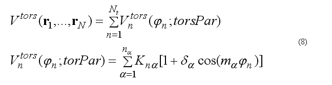
where torsion energy for bond j-k can have several torsion barriers with different multiplicity.
Torsion angle N is defined as
The forces on atoms i,j,k,l due to the single term of eq.(8b) are
The torsion energy and force are calculated via
subroutine torsanglenf(xyz1,xyz2,xyz3,xyz4,nTorsH,
& torsPar,eTors,f1,f2,f3,f4)
c torsPar(4*nTorsH) = {pass,Vt/2/pass,cos(delta),nFi },�
c eTors = sum{ Ki*[1+cos(delti)cos(i*Ftors)] }; i=1,..,nTorsH
c
Torsion parameters are taken from the LibData = bsparBATV.dat
The extraction of the torsion parameters from LibData = bsparBATV.dat for all quartets is done by
       subroutine getTorsPar(ffParFile,
     &              natom,atomNameEx,ResName,chName,ffAtomName,
     &              quar1234L,nQuar1234,quar1234Par,quar1234nPar)
c
c InPut:
c       ffParFile - ffParameters file
c       natom,atomNameEx,ResName,chName : PDB info
c       ffAtomName(ia) - FFatomName to search table
c       the quar1234L(i),i=1,..,nQuar1234 : the QuartetList
c RESULT: quar1234Par(16*nQuar1234) - torsionFF parameters for list
c         of  quartets
c         pass,Vt/2,delta,nFi - (printed) for each torsHarmonics,
c         pass,Vt/2/pass,cos(delta),nFi - finally in array
c        4- torsionHarmanics is possible.
c        quar1234nPar(iQuart) - number of torsHarmonics for the torsAngl
c
The improper torsion angle deformation keeps the four atoms 1-2-3-4 (i-j-k-l ) in specified geometry. The first atom in the improper quartet is a planar or (tetrahedral) atom. For example atoms Ci-CAi-N(i+1)-Oi are kept planar. The out of plane potential
CA-N-C-CB are kept in the tetrahedral configuration (L-amino acid) or CA-C-N-CB (D-amino acid) if CA in the united atom (CH) presentation.
The out of plane angle is defined for j-i-k four atoms with i is the planar (tetrahedral)
L
angle between to planes (i-j-k) and (j-k-l) with rotation angle around j-k, other words the
torsion angle in the sequence i-j-k-l
where
The forces on atoms i,j,kl due to a single term Vn
finally from the third Newton law
The improper energy and forces for a given improper quartet of atoms are calculated by the subroutine
c improper torsion energy force
c
        subroutine imprtorsanglenf(xyz1,xyz2,xyz3,xyz4,impPar,
     &                         eImpt,f1,f2,f3,f4)

c
c ImptPar(2) = K1, ksi0
All valence back-bond deformation are calculated in the file initAllForce.f

subroutine initAllForce(fcall,atomXYZ,
     &              makeVdWs,makeCLs,makeSLs,
     &              eVbondDef,vbdefForce,
     &              eVangDef,vAngdefForce,
     &              eImpDef,impDefForce,
     &              eTorsDef,torsAngForce,
     &              engVDWR1,vdwForceR1,
     &              engCOULR1,coulForceR1,
     &              engCOULR2,coulForceR2,
     &              restr1Eng,restr1AtForce,
     &              molSolEn, atomSolEn, atomSolFr)
c
        include 'xyzPDBsize.h'
        include 'xyzPDBinfo.h'
        include 'pair1234array.h'
        include 'nbondPairVCS.h'
        include 'vdw12Par.h'
        include 'restrainInfo.h'
        include 'loopInfo.h'
        include 'movingAtom.h'
        include 'solvGSarray.h'
        include 'optionPar.h'
c
 . . . . . . . . . . . . . . . . . . . . .

c
c all GeoDef forces are calculated at each step

 	call allAtVBondEForce(atomXYZ,
     &           natom,bond12List,nbond12,bond12ParL,
     &           eVbondDef,vbdefForce )
c
c
	call allAtVangEForce(atomXYZ,
     &           natom,trip123List,nTrip123,ang123ParL,
     &           eVangDef,vAngdefForce )
c
c
	call allAtImpTEForce(atomXYZ,
     &           natom,quarImp1234L,nImp1234,impAng1234ParL,
     &           eImpDef,impDefForce )
c
c torsionEnForces
c
        call allAtTorsEForce(atomXYZ,
     &           natom,quar1234List,nQuar1234,
     &           quar1234ParL,quar1234nPar,
     &           eTorsDef,torsAngForce )
c

 . . . . . . . . . . . . . . . . . . . . . . . . . . . . . . . . . . . . . . . . . . . . . . . . . . . . . . . . . .
The deformation forces are calculated at each time step in the MD run.
The non bonded pair interactions are calculated for the pair list. Pair list for the central atom i is a sequence of atom numbers for atom within the radius R from the central atom. Three separate pair lists are calculated. The Van der Waals pair list(i) includes atom j if
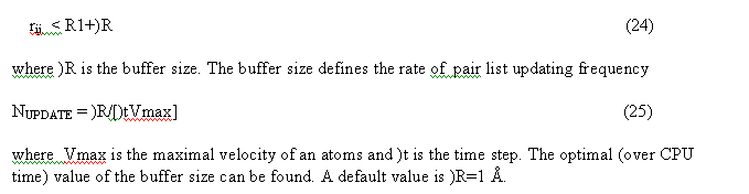
The linked list and all pairList (nnbPairLV, nnbPairLC, nnbPairLS) are calculated in the subroutine
c
        subroutine nonbondListVCS(rcutV,rcutC,rcutS,atomXYZ,atomQ,
     &           rbuffV,rbuffC,rbuffS,
     &           makeVdW,makeCL,makeS,
     &           natom,atomNumb,atomName,resName,chName,resNumb,
     &           nres,resNameRes,chNameRes,
     &           atomNameEx,startAtInRes,
     &           nmoveatom,moveAtomList,moveFlag,
     &           pair12List,startPairL12,nPairL12,
     &           pair13List,startPairL13,nPairL13,
     &           pair14List,startPairL14,nPairL14,
     &           nbpairListV,startnbPairLV,nnbPairLV,nnbpLVMAX,
     &           nbpairListC,startnbPairLC,nnbPairLC,nnbpLCMAX,
     &           nbpairListS,startnbPairLS,nnbPairLS,nnbpLSMAX)
c
fragment of code for the linked list calculation:
c distribute atoms over cells
c make linked list of atoms in cells
c headat(n) - head(incellN)
c linkList(ia) - linkedList
        ixm=1
        iym=1
        izm=1
        do ia = 1,natom
c calculate cell numb
        i3=3*ia-3
        xyzi(1)=atomXYZ(i3+1)-xMIN(1)
        xyzi(2)=atomXYZ(i3+2)-xMIN(2)
        xyzi(3)=atomXYZ(i3+3)-xMIN(3)
        ix = xyzi(1)/cellh+1
        iy = xyzi(2)/cellh+1
        iz = xyzi(3)/cellh+1
        if(ixm .lt. ix)ixm = ix
        if(iym .lt. iy)iym = iy
        if(izm .lt. iz)izm = iz
c cell number
        ncell = ix + (iy-1)*nsiz(1) + (iz-1)*nsiz(1)*nsiz(2)
        if(ncell .gt. ncell3MAX)then
        write(kanalp,*)'ERROR!:nonbondList: ncell3MAX is low !!'
        stop
        end if!

c make linked list
        linkList(ia) =  headat(ncell)
        headat(ncell) = ia
        end do !ia
c end of linked list calculation

The pair lists VDW and COULOMbic energy exclude 12, 13, 14 covalent bonded pairs. The Solvent model pairList
include all 12,13, 14 pairs.
The pair list are calculated for the range respectively:
c
        rcutV2 = (rcutV + rbuffV)**2     ! range for List1 -
                                                     VDWaals -  nbPairListV
        rcutV2m = (rcutV - rbuffC)**2    ! range for List2 - Coulombic twin
                                                        range - nbPairListC

        rcutC2p = (rcutC + rbuffC)**2    ! range for List2
        rcutS2 = (rcutS + rbuffS)**2     ! range for SolvationGSList -
                                                                nbPairListS
c

see file nonbobdListVCS.f
Van der waals forces are calculated for the non-bonded pair list nbpairListV()for atoms j within rij < RCUTV the cutoff radius for van der waals interactions. The modified potential 6-12 are used
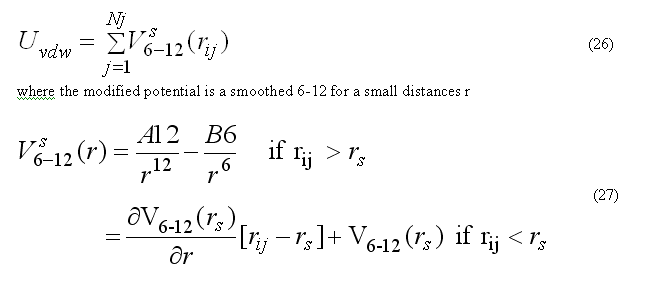
the pair list for atom i includes atoms j > i, to count each pair interaction once. The force Fvdwi on atom i due to interaction with atoms in the pair list
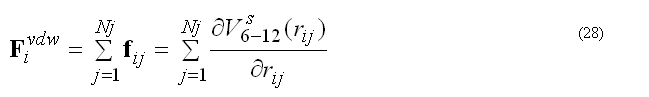
The modified (smoothed) 6-12 potential prevents over-flow when atoms are too close and generates smooth driving forces to resolve clash problems between atoms in molecular dynamics simulations, see
c
	subroutine vdwenforceij(dij2,dij1,rij,A12,B12,evdw,fi)
c
The coulombic energy and forces for atom i are calculated for all pairs within the radius RCUTC.
The coulombic energy/forces for a central atom i are calculated for the classical coulombic law or as a coulombic interaction between two charges on the compensating background charge uniformly distributed within the sphere of radius RCUTC
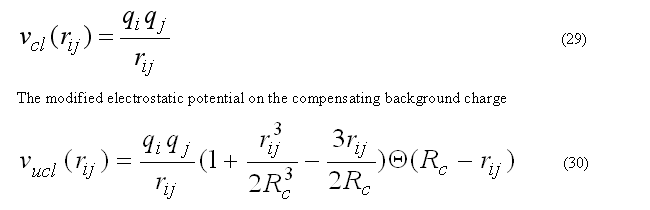
has zero interaction energy and forces for the rij > RCUTC. This form of electrostatic interactions is better suitable to prevent energy conservation in the molecular dynamic calculation, see
c
        subroutine coulenforceij(var,rcutC,dij2,dij1,rij,qi,qj,ecoul,fi)
c
The nonbonded energy and force within short range RCUTV=R1 are calculated in the subroutine
c allAtNonBondEForce : VDW and COULOMBIC
c
        subroutine allAtVDWEForceR1(atomXYZ,atomQ,
     &           natom,nmoveatom,moveAtomList,
     &           nbpairListV,startnbPairLV,nnbPairLV,
     &           pair14List,startPairL14,nPairL14,
     &           nVDWtype,atomVDWtype,atomVDW12ab,
     &           rcutV,rcutC,engVDW,vdwForce,engCOULR1,coulForceR1)
c
for the pair list nbpairListV() and pair14List(). The last one includes all 1-4 neihgbours
for which the amber force field uses the scaling factors for van der waals and coulombic interactions.
To increase performance of the van der waals energy/force calculations the table of
coefficient A12, B12 for all atom types are precalculated and then right values A12/B12 for a given
atom types in the pair ij are extracted from the vdw AB-parameter table
c get pointer to the AB table
        call vdw12TablePos(nVDWtype,t1,t2,t12)
        p4 = 4*t12
        A12 = atomVDW12ab(p4-3)
        B12 = atomVDW12ab(p4-2)
c
The long-range electrostatic forces within RCUTV < rij < RCUTC are calculated via the subroutine
c
        subroutine allAtVDWEForceR2(atomXYZ,atomQ,
     &           natom,nmoveatom,moveAtomList,
     &           nbpairListC,startnbPairLC,nnbPairLC,
     &           rcutR1,rcutR2,engCOULR2,coulForceR2)
c
c LongRamge -   RCUT1 < rij < RCUT2
The program keep separately the short-range and the long-range electrostatic energy and force.
The implicit solvation model - the Gaussian Shell model of Lazaridis & Karplus is used to calculate the solvation energy [POTEINS 35: 133-152, 1999]. The solvation free energy of the atom i
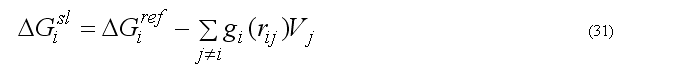
where sum is going over all neighbors of atom i which exclude volume Vj from the solvation volume around of the atom i. The function gi(r) describe the solvation energy density in the volume around the atom i and is approximated by the Gaussian function
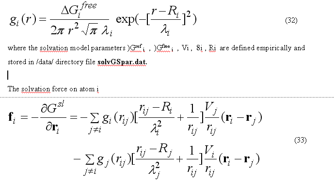
The sum over all solvation forces fi is zero.
The solvation forces are calculated by subroutine
c
	         call SolventEnForces(natom, atomXYZ,
     &         atomName,startPairL12,nPairL12,pair12List,
     &         nbpairListS,startnbPairLS,nnbPairLS,
     &         atomSolPar, molSolEn, atomSolEn, atomSolFr)
c
An MD run is performed by subroutine
c
	subroutine mdRun(ntimeMX,ntime0,ntime,ntimeR1,ntimeR2,
     &                ntimeF1,ntimeF2,ntimeF3,deltat,
     &                tempTg,tauTRF,atype,optra,wtra,nwtra,cltra)
c
c MD RUN propagates MDtraj from files in mdAtomXYZvel.h
c                                     [ atomXYZ0(*),atomVel0(*) ]
c    call initMDStart(T) inits the MD start
c                          from the INput atomXYZ(*)-->atom0XYZ(*)
c
c ntimeMX max number of time steps
c ntime0 - executed number of timesteps in the previous call
c ntime   executed number of timesteps in this call
c ntimeR1, ntimeR2 - update frequency  for R1, R2 pairLists
c ntimeF1,ntimeF2 - update freq for R1=(vdw+coulR1), R2-coulR2 en/forces
c ntimeF3 -  SOLVation forces
c GeoEn/force ntimeFg=1 - standart
c deltat- timestep, temp - initial(temp) of MD run
c tempTg - target T for NTV ansemble[K]
c tauTRF - tau Relaxation Factor [ps]
c atype - ansamble type = 0/1 - NEV, NTV
The MD algorithm consist of a long loop over the time steps.
For each time step MD trajectory is propagated for the )t = 1-2 femto sec,
as defined by user.
The pair lists are updated for each n-th timestep equal to ntimeR1, ntimeR2 for the short-range and for the twin-range long-range electrostatic energy calculations.
c
        call initNonBondList(atomXYZ0,makeVdW,makeCL,makeSL)
c
The atomic forces due to deformation of covalent structure and short-range non-bonded
calculation are updated for the each ntimeF1-th time step, the long-range
electrostatic are updated for the each ntimeF2-th step and solvation forces are updated for each ntimeF3-th time step.
{Note! In the current version the multiple time
step for pair list update and md equation integration are equal. The general case is not tested !}
c update forces/energy
	call initAllForce(fcall,atomXYZ0,doVdWef,doCLef,doSLef,
     &              eVbondDef,vbdefForce,
     &              eVangDef,vAngdefForce,
     &              eImpDef,impDefForce,
     &              eTorsDef,torsAngForce,
     &              engVDWR1,vdwForceR1,
     &              engCOULR1,coulForceR1,
     &              engCOULR2,coulForceR2,
     &              restr1Eng,restr1AtForce,
     &              molSolEn, atomSolEn, atomSolFr)
MD simulation can be done with a specified set of forces. The set of forces can be specified by the array fEngWF(*)
c
          eGeoDef   = fEngWF(1)*eVbondDef + fEngWF(2)*eVangDef
     &                + fEngWF(3)*eImpDef + fEngWF(4)*eTorsDef
     &                + fEngWF(8)* restr1Eng
          engCOUL   = fEngWF(6)*engCOULR1 + fEngWF(7)*engCOULR2
          engPOTENT = eGeoDef + fEngWF(5)*engVDWR1 + engCOUL +
     &                molSolEn*fEngWF(9)
c
For one time step propagation of the MD trajectory is done by the subroutine
c make mdStep
	call mdTimeStepProp(nmoveatom,moveAtomList,deltat)
c
which uses multi step leap-frog algorithm to calculate velocities and positions at time (t+deltat).
At each time step the temperature control routine performs calculation of the total kinetic energy
of the moving atoms. The relaxation the average temperature of the atomic system
to the specified value are give via the weak-coupling method or Berendsen method,
which scale the velocity by the factor lambTR(t)
Trajectory is written for each nwtra time steps. The trajectory can be written for atomic positions (and for atomic velocietis) in the user specified file.
Method description
Docking method is performed by subroutine runLigDock02 in the mdyn07 program procedure runLigDock02 perform ab initio docking of molecular ligand of size up to ~100 atoms.
The algorithm flow can be described as:
1) Calculation of the accessible surface of the protein. Calculation of a surface grid for probe sphere of radius ~ average atomic radius, and contact positions [bindSiteAt01(*)]with protein atoms. Calculation are done by subroutine surf_SAS04.
2) Calculation of a surface grid points for a probe ligand of radius of typical aromatic
ring [benzene] gridsizeSAS ~ 3.0 A. The surface grid are calculated by clustering of surface
contact positions bindSiteAt01(*) and the surface grid bindGridXYZSAS01(*) is generated. The contact
score [nsasGridPoint(*)] equal to the number of contact atomic positions included in to the surface
grid point bindGridXYZSAS01(*) is calculated.
The bindGridXYZSAS01(*) are sorted by descent of the contact score value
nsasGridPoint(*) and presents an initial trial positions for refined docking of ligand.
3) Refined docking is performed via subroutine runLigDock01(ig,bindGridXYZSAS01loc). For each initial positions bindGridXYZSAS01(*) for ligand center.
Procedure runLigDock01 perform global optimization of ligand orientation and position in a restrained region of 3D-space. Spatial restraints are a sphere of radius equal to gridsizeSAS. Orientational optimization based on exhaustive search via optimization from different initial orientations uniformly covering all orientational space. The orientational optimization can be done in two mode. Coarse grain mode consist of 24 orientations with 90deg between two neighbor orientations, fine mode consist of 144 orientations with 45deg angle between two neighbor orientations. For each initial ligand orientation the molecular dynamic simulated annealing coupled with van der waals potential scaling is performed for flexible ligand and fixed protein atoms. A variant of deformable potential energy surface global optimization method is used. Three best final position/orientations of ligand are collected for each initial positions bindGridXYZSAS01(*) in the files LigDockFinMMM.nnn.pdb - where MMM - grid position number, nnn - 001,002,003 - orientations.
The best docking variant for the ligand can be chosen as a file LigDockFinMMM.nnn.pdb with minimal potential energy engPOTENTLG:
Examples
1bty : benzamidine-trypsine complex
File #LigBindGridOnSAS: X Y Z contactScore ATOM 1 LBSt 1 16.536 26.130 8.764 11 ATOM 2 LBSt 2 29.319 14.972 16.378 11 ATOM 3 LBSt 3 6.595 15.454 32.366 9 ATOM 4 LBSt 4 28.049 26.396 3.572 9 ATOM 5 LBSt 5 37.370 14.662 29.278 8 ATOM 6 LBSt 6 9.605 28.662 39.481 7 ATOM 7 LBSt 7 18.280 35.574 15.402 7 ATOM 8 LBSt 8 30.648 34.679 44.060 7 ATOM 9 LBSt 9 34.040 33.767 21.484 7 ATOM 10 LBSt 10 5.056 19.922 18.987 6 ATOM 11 LBSt 11 25.308 5.865 13.437 6 ATOM 12 LBSt 12 13.241 31.812 30.019 6 ATOM 13 LBSt 13 6.174 15.317 15.623 6 ATOM 14 LBSt 14 15.230 11.995 39.322 6 ATOM 15 LBSt 15 42.858 27.966 33.933 6 ATOM 16 LBSt 16 39.046 14.805 5.421 5 ATOM 17 LBSt 17 24.676 37.002 14.221 5 ATOM 18 LBSt 18 39.100 25.116 6.122 5 ATOM 19 LBSt 19 25.156 6.498 5.813 5 ATOM 20 LBSt 20 14.736 13.757 2.279 5 ATOM 21 LBSt 21 35.933 31.703 11.547 5 ATOM 22 LBSt 22 45.035 21.844 22.099 5 ATOM 23 LBSt 23 12.210 8.874 28.161 5 ATOM 24 LBSt 24 11.197 11.080 32.573 5 ATOM 25 LBSt 25 25.549 16.554 -0.897 4 ATOM 26 LBSt 26 34.793 8.348 15.236 4 ATOM 27 LBSt 27 26.857 9.202 21.336 4 ATOM 28 LBSt 28 34.072 12.246 27.335 4 �..
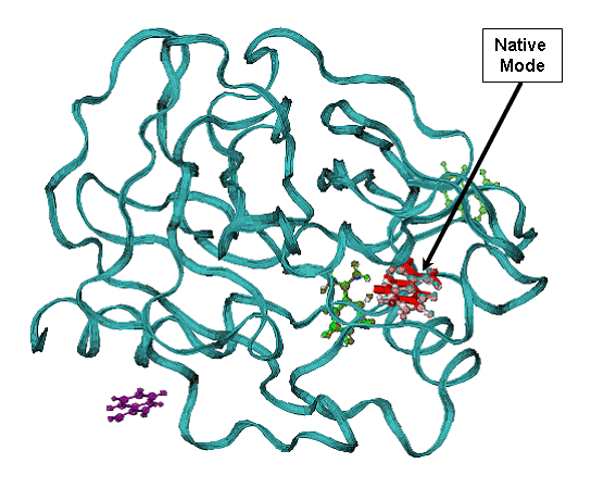
Fig.1. Docking results for benzamidine on trypsine - 1bty complex. |
A - contact Score (black square) for binding grid points vs refined potential energy of ligand binding (red diamonds).
|
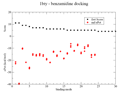 |
B - minimum energy docking mode (red bonds), RMSD = 0.54 A for all non Hydrogen
atoms ligand of the native binding mode.
CPK- green and violet are less favorable binding modes with low binding energy are shown in (A). CPK (pink) - native binding mode of benzamidine in 1bty. |
|
Fig.1. Docking results for benzamidine on trypsine - 1bty complex. |
A - contact Score (black square) for binding grid points vs refined potential energy of ligand binding (red diamonds).
|
|
B - minimum energy docking mode (red bonds), RMSD = 0.54 A for all non Hydrogen
atoms ligand of the native binding mode.
CPK- green and violet are less favorable binding modes with low binding energy are shown in (A). CPK (pink) - native binding mode of benzamidine in 1bty. |
|
|
Fig.2 Docking results for benzamidine on thrombin. |
A - Contact Score (black square) for binding grid points vs refined potential energy of ligand binding (red diamonds).
|
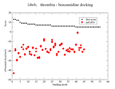 |
B(CPK blue) - minimum energy docking mode. Less favorable binding modes are shown - yellow, brown, green. CPK- (red) native benzamidine binding mode in 1dwb complex,
|
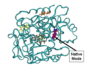
Minimum energy mode has RMSD = 0.27 A from the native binding mode of benzamidine. |
Fig.3. Docking result for biotine on streptavidine , 1stp complex. |
A - contact Score (black square) for binding grid points vs refined potential energy of ligand binding (red diamonds).
|
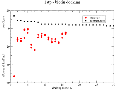 |
B - minimum energy docking mode structure of biotine - CPK, lines - native biotine in the 1stp complex.
|
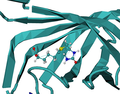
Minimum energy mode has RMSD = 0.96 A from the native binding mode of biotine. |
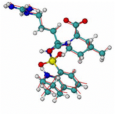
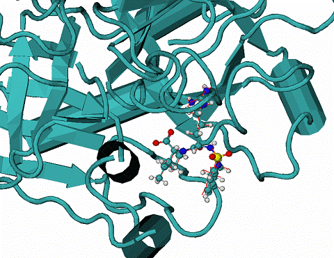
Fig. 4. Docking result for ILE-VAL dipeptide on Trypsinogen/pancreatic trypsin inhibitor. |
A - contact Score (black square) for binding grid points vs refined potential energy of ligand binding (red diamonds).
|
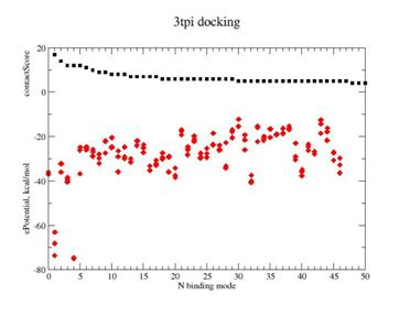 |
B - Lines are minimum energy docking modes of rank 1- 4 structures of ILE-VAL peptide - lines, CPK - native binding mode of biotine in the 1stp complex.
|
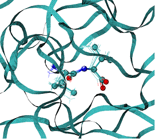
The best binding energy mode has RMSD = 0.46 A from the native binding mode of dipeptide ILE-VAL |
Table 1. Energies of top ranked binding modes, and RMSD from the native binding mode.
| Binding mode | ePL, kcal/mole | RMSD, A |
| Rank 1 -
LigDockFin001.001.pdb |
-76.07 | 0.46 |
| Rank2 -
LigDockFin001.002.pdb |
-75.6 | 0.58 |
| Rank3 -
LigDockFin001.002.pdb |
75.5 | 0.78 |
| Rank4 -
LigDockFin004.001.pdb |
-74.8 | 0.88 |
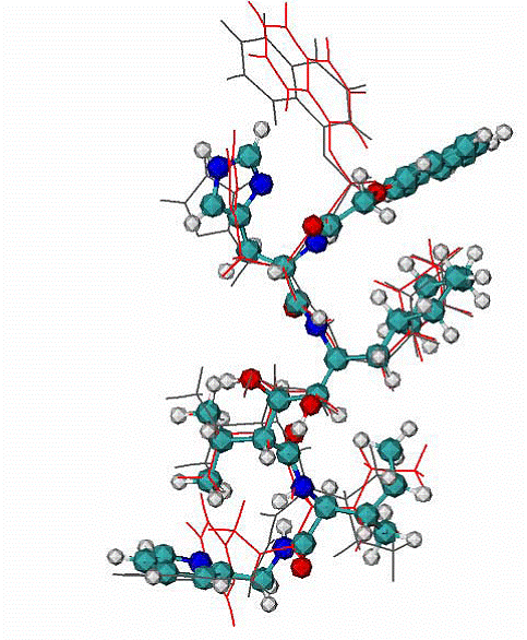
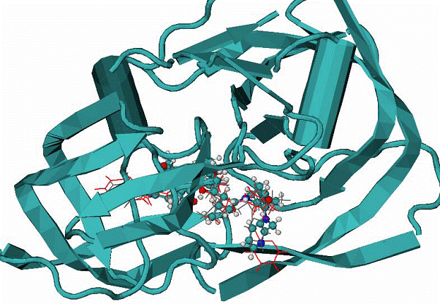
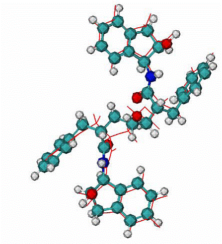
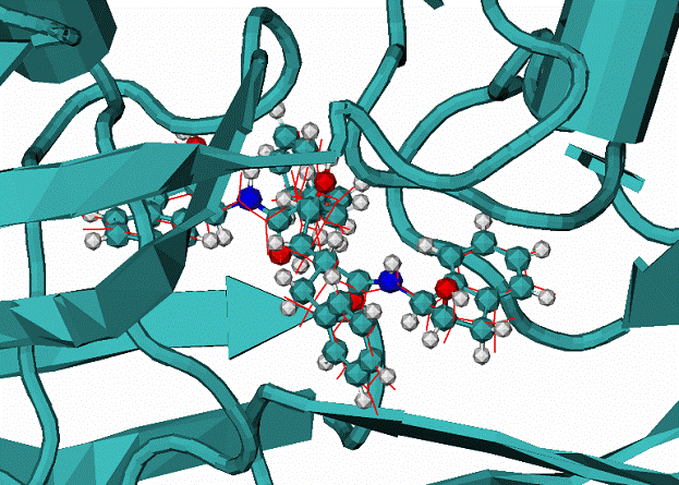
Fig. 5. 1dwc complex of Human thrombin with thrombin-inhibitor MIT . |
Human thrombin - 296 residues;
MIT - molecule includes 80 atoms |
 |
|
Fig. 6. 1hiv complex of HIV1 protease with inhibitor NOA
|
A -
Two top ranked calculated binding modes of NOA in comparison with the NOA ligand in the native binding mode of 1hiv complex. CPK - native binding mode, lines (red and grey) the top ranked mode by energy of binding. The RMSD from the native are ~3.1A for all atoms. The major difference between native and calculated modes are the orientation of one aromatic double-ring at the top of molecule NOA, the RMSD = 1.1. A over all atoms except the later aromatic system.
|
 |
B - 1hiv complex of HIV1 protease with inhibitor NOA CPK - native mode, red and grey lines - are calculated modes. |
|
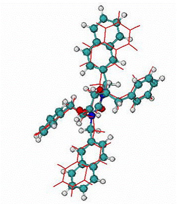
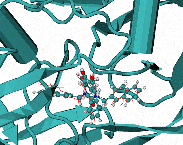
Fig. 7. 1hvr complex of HIV1 protease with inhibitor XK2
|
A -
Calculated binding mode of XK2, red lines, CPK - native binding mode of XK2 ligand. RMSD = 0.95 A for all atom.
|
 |
B - Calculated docking mode for the ligand XK2 in complex with HIV1 protease, CPK - the native binding mode of the XK2 ligand.
|
|
Fig. 8. 1hvp complex of 1HIV protease with VAC molecule inhibitor
|
A -
Calculated best binding mode of VAC is in red lines, CPK - native VAC inhibitor in the 1hvp complex; the RMSD = 0.99 A.
|
 |
B - 4hvp complex, red lines is the calculated mode, CPK - the native binding mode of VAC inhibitor.
|
|
| Table 1. Results of MdDock method for a set of complexes | |||
| complex | Ntors | RMSD, A | DEgap |
| 1) 1bty trypsin/benz | 0 | 0.5 | 9.7 |
| 2) 1dwb a-thrombin/benz | 0 | 0.5 | 13.3 |
| 3) 1stp streptavidine/biotin | 5 | 0.96 | 29.5 |
| 4) 3tpi trypsinogen/Ile-Vla | 6 | 0.42 | 10.6 |
| 5) 1dwc a-thrombin/MIT | 8 | 0.2 | 10.8 |
| 6) 1hiv HIV1 protease/NOA | 16 | 1.1/3.1 | 2.6 |
| 7) 1hvr HIV1 protease/XK263 | 8 | 0.95 | 39.1 |
| 8) 4phv HIV1 protease/VAC | 15 | 0.9 | 3.4 |
Ntors - number of flexible torsion angles.
DEgap - energy gap between lowest energy binding mod and the next energy mode.
Conclusion:
The developed method of blind docking has show a good accuracy in prediction of the native bindig modes of flexible ligands. At the test set of 8 ligands the method shows 100% accuracy, i.e. the native binding mode are found as the mode with highest binding affinity.
Tamar Schlick. Molecular Modeling and simulation. Springer-Verlag, New York, 2000.
Cornell W.D., Cieplak P., Bayly C.I., Gould I.R., Mertz K.M.,
Ferguson D., Spellmeyer D.C., Fox T., Caldwell J.W., Kollam P.A.
A second generation force field for the simulation of proteins, nucleic acids and organic molecules.
J.Am.Chem.Soc. 1995: 117, p.5179-5197
Lazaridis T., Karplus M. Proteins: Structu, Funct., and Gen. 1999: 35, p.133-152